Home
Uncategories
Arteries In Neck Diagram / Arteries In The Neck Picture - Human Anatomy Body - Learn vocabulary, terms and more with flashcards, games and other study tools.
Arteries In Neck Diagram / Arteries In The Neck Picture - Human Anatomy Body - Learn vocabulary, terms and more with flashcards, games and other study tools.
Arteries In Neck Diagram / Arteries In The Neck Picture - Human Anatomy Body - Learn vocabulary, terms and more with flashcards, games and other study tools.. Ploaded with beautifully illustrated diagrams clearly and concisely labeled for easy identification. They originate from the carotid bifurcation, travel through the. Rarely artery ascends in the neck without undergoing division, either eca or ica being absent. Origin the right common carotid artery originates behind the sternoclavicular. The axillary artery represents the continuation of the subclavian artery and is a major artery of the upper limb.
Each side of the neck contains two triangular sections created by the major deep muscles. The anatomy of the neck, part three: It commences in the substance of the parotid gland, on a level with the angle of the mandible, and runs perpendicularly down the neck, in the direction of a line drawn from the angle of the mandible to the middle of the clavicle at the posterior border of the sternocleidomastoideus. Rarely artery ascends in the neck without undergoing division, either eca or ica being absent. Ploaded with beautifully illustrated diagrams clearly and concisely labeled for easy identification.
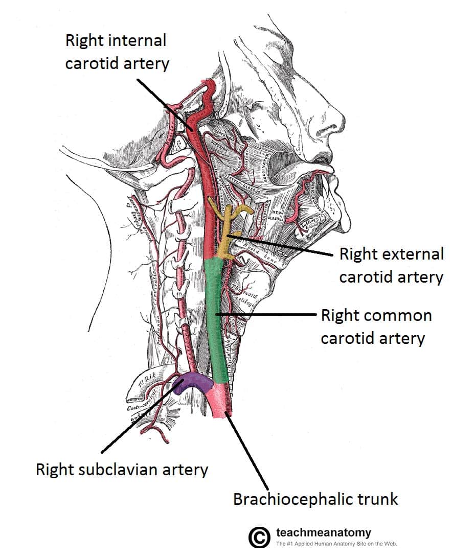
Blood Vessels and Lymphatics of the Head and Neck - TeachMeAnatomy from teachmeanatomy.info In the following diagrams, the anatomy is drawn with and without the labels. Muscles of the neck and their blood supply these pictures of this page are about:arteries in your neck. Posted by cassidy smith on 9 may 2018, 11:14 am. Start studying arteries in the neck. Schematic owchart from the arteries in the neck and head. Instant anatomy is a specialised web site for you to learn all about human anatomy of the body with diagrams, podcasts and revision questions. The distal part of the subclavian artery can be located as it emerges between the anterior and middle scalene muscles. They originate from the carotid bifurcation, travel through the.
The sternocleidomastoid muscle separates the sections, known as the anterior and posterior triangles.
Rarely artery ascends in the neck without undergoing division, either eca or ica being absent. You can use highlighters in different colors to see which notes are in a major scale, a minor scale, or a. The thyroid gland is located in the neck below the thyroid cartilage, or adam's apple. It is extremely important because every cell in the body depends on the hormones the thyroid produces to determine how quickly to convert calories and ox. Instant anatomy is a specialised web site for you to learn all about human anatomy of the body with diagrams, podcasts and revision questions. Continuation of the subclavian artery as it passes under the midpoint of the clavicle on the outer edge of the fir. The left and right carotids, and the left and right vertebral arteries. The neck diagram above shows you the structure and anatomy of the neck. A person with neck swelling has enlargement of the soft tissues that covers the neck. The anatomy of the neck, part three: Brain diagram brain anatomy anatomy and physiology human anatomy carotid artery human anatomy picture definition conditions more. The internal carotid artery is a major branch of the common carotid artery, supplying several parts of the head with blood, the most important one being the brain. There are 2 common carotid arteries:
Start studying arteries in the neck. The carotids reside beneath the skin on either side, and the pulse can be felt easily with your hand. A blockage in one of the carotid arteries can be cleared either by endarterectomy or carotid angioplasty. The distal part of the subclavian artery can be located as it emerges between the anterior and middle scalene muscles. Brain diagram brain anatomy anatomy and physiology human anatomy carotid artery human anatomy picture definition conditions more.
Useful Notes on the Internal Jugular Vein of Human Neck | Human Anatomy from cdn.yourarticlelibrary.com Schematic owchart from the arteries in the neck and head. The sternocleidomastoid muscle separates the sections, known as the anterior and posterior triangles. You can use highlighters in different colors to see which notes are in a major scale, a minor scale, or a. Thirdly, blank neck diagrams are useful in mapping out scales. Ploaded with beautifully illustrated diagrams clearly and concisely labeled for easy identification. The external carotid artery reduces in size while moving up the neck, giving various branches along the way. The neck arteries 3 pages 1918 human anatomy by mysunshinevintage. A blockage in one of the carotid arteries can be cleared either by endarterectomy or carotid angioplasty.
Posted by cassidy smith on 9 may 2018, 11:14 am.
There are two internal carotid arteries in total, one on each side of the neck. Thirdly, blank neck diagrams are useful in mapping out scales. They can be called the main arteries of the head and neck. Smartdraw includes 1000s of professional healthcare and anatomy head & neck lateral view of the head with arteries of the head and neck shown in relation to underlying skeletal structures. Discover the power of the scale generator in neck diagrams pro! It is extremely important because every cell in the body depends on the hormones the thyroid produces to determine how quickly to convert calories and ox. The external carotid artery supplies the areas of the head and neck external to the start studying arteries of head and neck. Rarely artery ascends in the neck without undergoing division, either eca or ica being absent. Arteria carotis interna) is located in the inner side of the neck in contrast to the external carotid artery. Each side of the neck contains two triangular sections created by the major deep muscles. As it crosses the first rib, it becomes the axillary artery, which goes onto supply the upper limb. Blank neck diagrams are important to musicians for several reasons. Continuation of the subclavian artery as it passes under the midpoint of the clavicle on the outer edge of the fir.
Posted by cassidy smith on 9 may 2018, 11:14 am. 20 5 circulatory pathways anatomy and physiology. The distal part of the subclavian artery can be located as it emerges between the anterior and middle scalene muscles. As it crosses the first rib, it becomes the axillary artery, which goes onto supply the upper limb. The external carotid artery reduces in size while moving up the neck, giving various branches along the way.
Teach Besides Me: Pictures Of Veins And Arteries from o.quizlet.com Schematic owchart from the arteries in the neck and head. Neck diagram of muscles, arteries, and skeleton. Lateral view of the head with veins of the head and neck shown in relation to underlying skeletal structures. The thyroid gland is located in the neck below the thyroid cartilage, or adam's apple. The axillary artery represents the continuation of the subclavian artery and is a major artery of the upper limb. Blank neck diagrams are important to musicians for several reasons. The sternocleidomastoid muscle separates the sections, known as the anterior and posterior triangles. Brain diagram brain anatomy anatomy and physiology human anatomy carotid artery human anatomy picture definition conditions more.
The neck diagram above shows you the structure and anatomy of the neck.
The left and right carotids, and the left and right vertebral arteries. There are 2 common carotid arteries: A blockage in one of the carotid arteries can be cleared either by endarterectomy or carotid angioplasty. As it crosses the first rib, it becomes the axillary artery, which goes onto supply the upper limb. Blank neck diagrams are important to musicians for several reasons. Printable neck diagrams to help you learn more about the system that makes up our neck. The internal carotid artery (latin: Muscles of the neck and their blood supply these pictures of this page are about:arteries in your neck. Instant anatomy is a specialised web site for you to learn all about human anatomy of the body with diagrams podcasts and revision q. It commences in the substance of the parotid gland, on a level with the angle of the mandible, and runs perpendicularly down the neck, in the direction of a line drawn from the angle of the mandible to the middle of the clavicle at the posterior border of the sternocleidomastoideus. In the neck, the following diagram points out the major landmarks of the neck. Discover the power of the scale generator in neck diagrams pro! Arteria carotis interna) is located in the inner side of the neck in contrast to the external carotid artery.
There are 2 common carotid arteries: arteries in neck. Thirdly, blank neck diagrams are useful in mapping out scales.


0 Comments:
Post a Comment